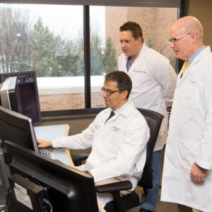
Nuclear medicine refers to a category of diagnostic imaging that uses small amounts of radioactive substances to obtain images of various functions inside the body in order to diagnose or determine the severity of cancer, heart disease, gastrointestinal, endocrine, neurological and other conditions or abnormalities. Common nuclear medicine tests include bone scans (including SPECT), bilary scans, cardiac MUGAs and thyroid imaging.
Nuclear Medicine

FAQS
Usually, tiny amounts of radioactive material are injected into the patient’s vein through a standard IV. Pictures are then taken with a Gamma camera (essentially a sophisticated Geiger counter) that measures how much radioactivity the area being examined is registering, ultimately generating images that reflect important functional anatomy.
The amount of radiation the body is exposed to is very small. It is actually smaller than many types of regular x-rays.
Gallbladder Nuclear Medicine Study (A HIDA Scan)
Please do not eat or drink anything for 6 hours before the study.
Other Nuclear Medicine Studies
If special preparation is necessary, we will explain it to you when you schedule your examination.
Our radiologists look at all of your pictures and compare this study with any previous examinations you may have had. Our typed report is available to your doctor usually within one day.
