X-Ray/Fluoroscopy

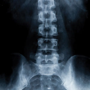
X-ray is the oldest and most frequently used form of imaging to see inside the human body. It uses a focused beam and a special detector to obtain images of body anatomy. It is also a safe and generally non-invasive procedure. Fluoroscopy is a special technique that uses x-rays to see the body’s internal structures and organs in motion.

X-rays are absorbed by different body tissues in varying degrees. Dense tissue, like bone, absorbs most x-rays and appears white on the image. Less dense tissues appear in shades of gray. X-rays that pass through air, like in the lungs and colon, aren’t absorbed at all and appear black on the image.
The x-ray is used for a variety of reasons when images of internal body structures are needed.

x-ray/fluoroscopy

FAQS

It depends on what part of the body is examined. For chest x-rays, the amount of radiation a patient receives is very tiny—only a fraction of the amount of radiation a person gets while walking around in the sunshine over a year. Although still quite small, the dosage may be larger for other types of x-rays. For this reason, you only want to have x-rays if you really need them.

No patient preparation is needed.

Our radiologists interpret all of your pictures and compare your study with any of your previous examinations. Our typed report is available to your doctor usually within one day.

We recommend reaching out to the facility it will be performed at for prep.

Please do not eat or drink anything on the morning of the examination. Even drinking a small amount of fluid on the morning of the examination is enough to prohibit adequate barium coating of your stomach, making the study of poor quality.

Skip to content